
Brain Labeled Diagram
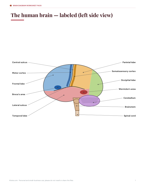
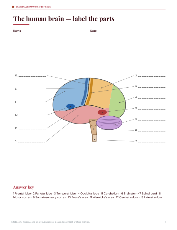
The human brain is divided into regions with different jobs. This labeled diagram shows the left side of the brain with its four lobes, the cerebellum and brainstem, the strips of motor and sensory cortex, and the two classic language areas. Click any region to see what it does, switch off the labels to test yourself, or try the quizzes — then print a labeled or blank copy.
- Free to use
- No sign-up
- Runs in your browser
Interactive brain diagram
Brain Diagram Worksheet Pack
Printable brain worksheets with answer keys: labeled diagram, blank numbered diagram, function matching, a lobes colouring sheet and a 12-question quiz.
- Labeled diagram (PDF)
- Blank diagram (PDF)
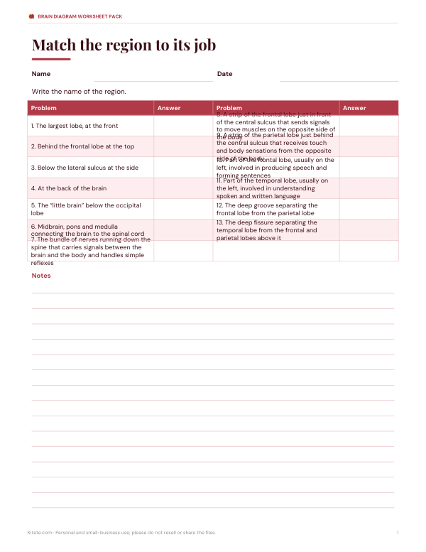
- Function matching (PDF, DOCX)
- 12-question quiz (PDF, DOCX)
Formats: PDF, DOCX. Instant download after payment (link valid 72 hours, up to 5 downloads). AI-assisted: the templates were drafted with AI help and reviewed and laid out before publishing.
$3.00 USD, one-time 4 files · about $0.75 each
- Instant download
- 7-day refund
- Secure checkout by Stripe
7-day money-back guarantee. Full refund within 7 days of purchase, even after you download.
Free sample: Brain Blank (PDF, 65 KB)
Full refund within 7 days — see the refund policy and license.
Free printables
Single sheets you can print right now, no sign-up. The full pack adds every sheet in one download plus the editable files.
 Printable Brain Diagram Quiz
Parts of the brain and what they do
| Region | Main functions |
|---|---|
| Frontal lobe | The largest lobe, at the front. It handles planning, decision-making, problem-solving, attention, personality and voluntary movement. |
| Parietal lobe | Behind the frontal lobe at the top. It combines touch, temperature, pain and body position, and helps with spatial awareness and numbers. |
| Temporal lobe | Below the lateral sulcus at the side. It processes hearing and language understanding and, with the hippocampus inside it, forming new memories. |
| Occipital lobe | At the back of the brain. It is the main visual processing centre, interpreting colour, shape and motion. |
| Cerebellum | The “little brain” below the occipital lobe. It coordinates balance, posture and smooth, precise movement and helps learn motor skills. |
| Brainstem | Midbrain, pons and medulla connecting the brain to the spinal cord. It controls automatic functions such as breathing, heart rate, swallowing and sleep–wake cycles. |
| Spinal cord | The bundle of nerves running down the spine that carries signals between the brain and the body and handles simple reflexes. |
| Motor cortex | A strip of the frontal lobe just in front of the central sulcus that sends signals to move muscles on the opposite side of the body. |
| Somatosensory cortex | A strip of the parietal lobe just behind the central sulcus that receives touch and body sensations from the opposite side of the body. |
| Broca’s area | Part of the frontal lobe, usually on the left, involved in producing speech and forming sentences. |
| Wernicke’s area | Part of the temporal lobe, usually on the left, involved in understanding spoken and written language. |
| Central sulcus | The deep groove separating the frontal lobe from the parietal lobe. |
| Lateral sulcus | The deep fissure separating the temporal lobe from the frontal and parietal lobes above it. |
The cerebrum and its four lobes
The cerebrum is the largest part of the brain, split into left and right hemispheres joined by a thick band of nerve fibres, the corpus callosum. Its wrinkled outer layer, the cerebral cortex, is only 2–4 mm thick but folded into ridges (gyri) and grooves (sulci) that pack a large surface into the skull. Two deep grooves divide each hemisphere into lobes: the central sulcus separates the frontal lobe from the parietal lobe, and the lateral sulcus separates the temporal lobe below. The occipital lobe sits at the back.
Left and right: crossed control
Each hemisphere mainly controls and senses the opposite side of the body — the left motor cortex moves the right hand. The two hemispheres also specialise to some degree: in most people, language areas such as Broca’s and Wernicke’s areas are in the left hemisphere, while the right hemisphere plays a larger part in some aspects of spatial attention and recognising faces. The popular idea of “left-brained” and “right-brained” personalities isn’t supported by research; both hemispheres work together in almost everything we do.
How to use the interactive diagram
- Start in Labeled mode and click each region to read what it does.
- Switch to Numbered mode to hide the names — print it as a worksheet.
- In “Spot the region”, one area is highlighted and you pick its name.
- In the function quiz, you read a description and choose the matching part.
- Print the labeled or numbered diagram for revision.
Worked example: tracing a sentence
Hearing a friend ask “Can you pass the salt?” involves several regions. The sound is processed first in the auditory cortex of the temporal lobe; Wernicke’s area helps interpret the meaning. Deciding to respond involves the frontal lobe, and Broca’s area helps form the words “Here you are.” To reach for the salt, the motor cortex sends signals to the arm while the parietal lobe tracks where the salt and your hand are, the occipital lobe processes what you see, and the cerebellum makes the movement smooth and accurate. The brainstem keeps you breathing throughout.
Motor and sensory maps
The motor cortex and somatosensory cortex each contain a map of the body, often drawn as a distorted figure called a homunculus. Parts of the body with fine control or sensitivity — the hands, lips and tongue — have much larger areas of cortex than the back or legs. This is why a small injury in one part of the motor cortex may affect the hand while leaving the leg unaffected.
What the diagram simplifies
This is a simplified side view. Real brains vary in shape, and many important structures are hidden inside or seen only from the middle: the corpus callosum, thalamus, hypothalamus, hippocampus, amygdala and the limbic system. Brain functions are also far more connected than a map of regions suggests — most tasks involve networks spread across several areas. Use the diagram to learn the main landmarks.
Memory tips
- Frontal = Future planning and Fine decisions.
- Parietal = Position and Physical sensation.
- Temporal = Talk (understanding) and Tunes (hearing), at the Temples.
- Occipital = Optics — vision at the back.
- Cerebellum = balance, like a little gyroscope under the back of the brain.
Frequently asked questions
What are the main parts of the brain?
The cerebrum (with frontal, parietal, temporal and occipital lobes), the cerebellum and the brainstem.
What does the frontal lobe do?
Planning, decision-making, personality and voluntary movement.
Which part of the brain controls balance?
The cerebellum.
Where is Broca’s area?
In the frontal lobe, usually on the left, near the lateral sulcus.
What does the brainstem control?
Automatic functions such as breathing, heart rate and swallowing.
Can I print a blank brain diagram?
Yes, choose Numbered mode and print.